Anatomy Of Chest Organs : Human Anatomy Chest Abdomen Anterior Posterior Vintage Medical : Navegue pelas 146 anatomy of the chest organs imagens e fotografias de stock disponíveis ou comece uma nova pesquisa para explorar mais imagens e fotografias de stock.
Anatomy Of Chest Organs : Human Anatomy Chest Abdomen Anterior Posterior Vintage Medical : Navegue pelas 146 anatomy of the chest organs imagens e fotografias de stock disponíveis ou comece uma nova pesquisa para explorar mais imagens e fotografias de stock.. An organ is a collection of tissues joined in a structural unit to serve a common function. Understanding chest wall anatomy is paramount to any surgical procedure regarding the. It describes the theatre of events. Poster showing anterior and posterior views of the heart, and left and right ventricles. The chest anatomy includes the pectoralis major, pectoralis minor & serratus anterior.
‒ topographic anatomy and operative surgery (main surgical approaches to abdominal and chest organs) test questions from related disciplines: The study of the anatomy of the chest is very important because the importance of the heart and lungs is seen. Poster showing anterior and posterior views of the heart, and left and right ventricles. Among the major organs contained in the thoracic cavity are the heart and lungs. Radiology basics of chest ct anatomy with annotated coronal images and scrollable axial images to help medical students and junior doctors learning anatomy.
Overview Of Chest Muscles from www.modernheal.com The study of the anatomy of the chest is very important because the importance of the heart and lungs is seen. ‒ human anatomy (localization and structure of internal organs); Human anatomy human internal organs dummy, training dummy, detail of the face, thorax and intestines. Diagrams showing the general organisation of the thorax with the pleural cavity. Anatomy is to physiology as geography is to history: Normal and topographic anatomy of abdominal organs. Among the major organs contained in the thoracic cavity are the heart and lungs. The chest itself is supported and protected by various muscles covering the ribcage, the spine, and shoulders.
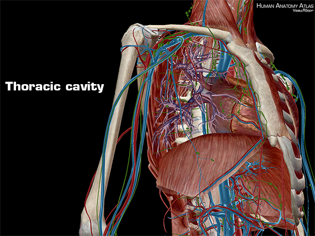
Central compartment (mediastinum),… thoracic cage (rib cage).
Find the perfect anatomy of the chest organs stock photos and editorial news pictures from getty images. Among the major organs contained in the thoracic cavity are the heart and lungs. The thorax or chest is a part of the anatomy of humans and various other animals located between the neck and the abdomen. Learn about each muscle, their locations & functional anatomy. Graphic shows the complex and diverse structures and organs of the thorax. Normal and topographic anatomy of abdominal organs. ‒ topographic anatomy and operative surgery (main surgical approaches to abdominal and chest organs) test questions from related disciplines: Human anatomy human internal organs dummy, training dummy, detail of the face, thorax and intestines. It contains organs including the heart, lungs, and thymus gland, as well as muscles and various other internal structures. Central compartment (mediastinum),… thoracic cage (rib cage). Selecione entre imagens premium de anatomy of the chest organs da mais elevada qualidade. .normal anatomy and confirm variants,imaging anatomy: ‒ human anatomy (localization and structure of internal organs);
Radiology basics of chest ct anatomy with annotated coronal images and scrollable axial images to help medical students and junior doctors learning anatomy. ‒ topographic anatomy and operative surgery (main surgical approaches to abdominal and chest organs) test questions from related disciplines: Chest scan showing a large hydropneumothorax from pleural empyema on the right side of the chest cavity (a is air; It provides access to ct images in the axial plane, allowing the user to learn and. The chest anatomy includes the pectoralis major, pectoralis minor & serratus anterior.
Anatomy and Physiology: Anatomical Planes and Cavities from www.visiblebody.com The thorax or chest is a part of the anatomy of humans and various other animals located between the neck and the abdomen. It contains organs including the heart, lungs, and thymus gland, as well as muscles and various other internal structures. The user can browse between different groups of images using the series tab: Find the perfect anatomy of the chest organs stock photos and editorial news pictures from getty images. Diagrams showing the general organisation of the thorax with the pleural cavity. Poster showing anterior and posterior views of the heart, and left and right ventricles. How to view the anatomical labels. The anatomical drawings were organized in a fairly classical manner to be easily used as a standard anatomical atlas.
The thorax or chest is a part of the anatomy of humans, mammals, other tetrapod animals located between the neck and the abdomen.
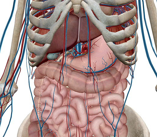
Anatomy is to physiology as geography is to history: The chest itself is supported and protected by various muscles covering the ribcage, the spine, and shoulders. .normal anatomy and confirm variants,imaging anatomy: It contains organs including the heart, lungs, and thymus gland, as well as muscles and various other internal structures. Find the perfect chest anatomy stock photo. The study of the anatomy of the chest is very important because the importance of the heart and lungs is seen. The sternum is located along the body's midline in the anterior thoracic region just deep to the skin. It describes the theatre of events. Navegue pelas 146 anatomy of the chest organs imagens e fotografias de stock disponíveis ou comece uma nova pesquisa para explorar mais imagens e fotografias de stock. Organs exist in most multicellular organisms, including not only humans and other animals but also plants. This atlas is a comprehensive and affordable learning tool for medical students and residents and especially for radiologists and pneumologists. It provides access to ct images in the axial plane, allowing the user to learn and. Chest, abdomen, pelvisprovides detailed views of anatomic structures in successive imaging chest wall muscle chest wall subcutaneous tissue pleura.
It describes the theatre of events. Where is the sternum found. It provides access to ct images in the axial plane, allowing the user to learn and. The heart beats around 100,000 times a day, pumping approximately 8 pints of blood throughout the body 24/7. Organs exist in most multicellular organisms, including not only humans and other animals but also plants.
Chest Cavity Anatomy - Anatomy Drawing Diagram from www.visiblebody.com Anatomy is to physiology as geography is to history: Human anatomy human internal organs dummy, training dummy, detail of the face, thorax and intestines. Radiology basics of chest ct anatomy with annotated coronal images and scrollable axial images to help medical students and junior doctors learning anatomy. Normal and topographic anatomy of abdominal organs. Showing the myriad different appearances of normal anatomic structures is beyond the scope of this chapter; Chest scan showing a large hydropneumothorax from pleural empyema on the right side of the chest cavity (a is air; Poster showing anterior and posterior views of the heart, and left and right ventricles. And flexibility to aid in the functional process of respiration.
They are learned by paying close attention to.
The study of the anatomy of the chest is very important because the importance of the heart and lungs is seen. The heart beats around 100,000 times a day, pumping approximately 8 pints of blood throughout the body 24/7. The chest itself is supported and protected by various muscles covering the ribcage, the spine, and shoulders. The chest or thorax is the region between the neck and diaphragm that encloses organs, such as the heart, lungs, esophagus, trachea, and thoracic diaphragm. Poster showing anterior and posterior views of the heart, and left and right ventricles. Anatomy is to physiology as geography is to history: Central compartment (mediastinum),… thoracic cage (rib cage). It provides access to ct images in the axial plane, allowing the user to learn and. Chest scan showing a large hydropneumothorax from pleural empyema on the right side of the chest cavity (a is air; Organs exist in most multicellular organisms, including not only humans and other animals but also plants. Diaphragm, thoracic nerve diagram, human anatomy, abdomen human body, chest human body, human thorax 3d, human body diagram appendix, human body diagram liver, human body diagram organs, human. Among the major organs contained in the thoracic cavity are the heart and lungs. Find the perfect chest anatomy stock photo.
The heart beats around 100,000 times a day, pumping approximately 8 pints of blood throughout the body 24/7 anatomy of chest. The heart beats around 100,000 times a day, pumping approximately 8 pints of blood throughout the body 24/7.


0 Komentar